What does an Alzheimer's amyloid clump look like? Research it in 3D
Amyloid-beta clumps build up between brain cells in Alzheimer's. You can measure how one clump is stacked in a free structure file, PDB 5OQV.
"My grandmother forgot my name before she passed." Swap in a grandfather or a great-aunt, and a lot of teenagers could say the same. It's a quiet kind of loss, and it raises a question that feels far too big for a high school student: can scientists find a way to stop Alzheimer's?
You won't answer that in one project. But a small, real piece of it is open to you now, on a laptop, with free data.
Alzheimer's is a disease, not normal aging
You've probably heard that forgetting things is just part of getting old. Some forgetfulness is, but dementia isn't, says the US National Institute on Aging (NIA). Alzheimer's is the most common cause of dementia in older adults, and NIA describes it as a brain disorder that slowly destroys memory and thinking skills.
In 1906, Dr. Alois Alzheimer examined the brain of a woman who had died of an unusual mental illness with memory loss and language problems. He found abnormal clumps, now called amyloid plaques, and tangled fibers, now called tau tangles. They're still considered main features of the disease.
Plaques are built from amyloid-beta, a small piece cut from a larger protein called the amyloid precursor protein. Amyloid-beta collects in the spaces between neurons (nerve cells), while tau tangles form inside them. NIA notes that one form, amyloid-beta 42, is thought to be especially toxic.
If you've wondered why this protein starts sticking to itself, you're asking a question researchers are still working on.
Families often notice Alzheimer's first as lost names and faces. Underneath, plaques build up between nerve cells and tangles form inside them.
How one drug strategy goes after the clumps
One approach: if amyloid-beta clumps help cause the damage, find them and remove them. In research, that takes three parts.
Choose which clump to target. Amyloid-beta can form small soluble (dissolved) clumps called oligomers or protofibrils, or long, insoluble fibrils that pile up into plaques.
Have a question you want to investigate?
Explore our faculty-guided programs, or tell admissions what you are curious about.
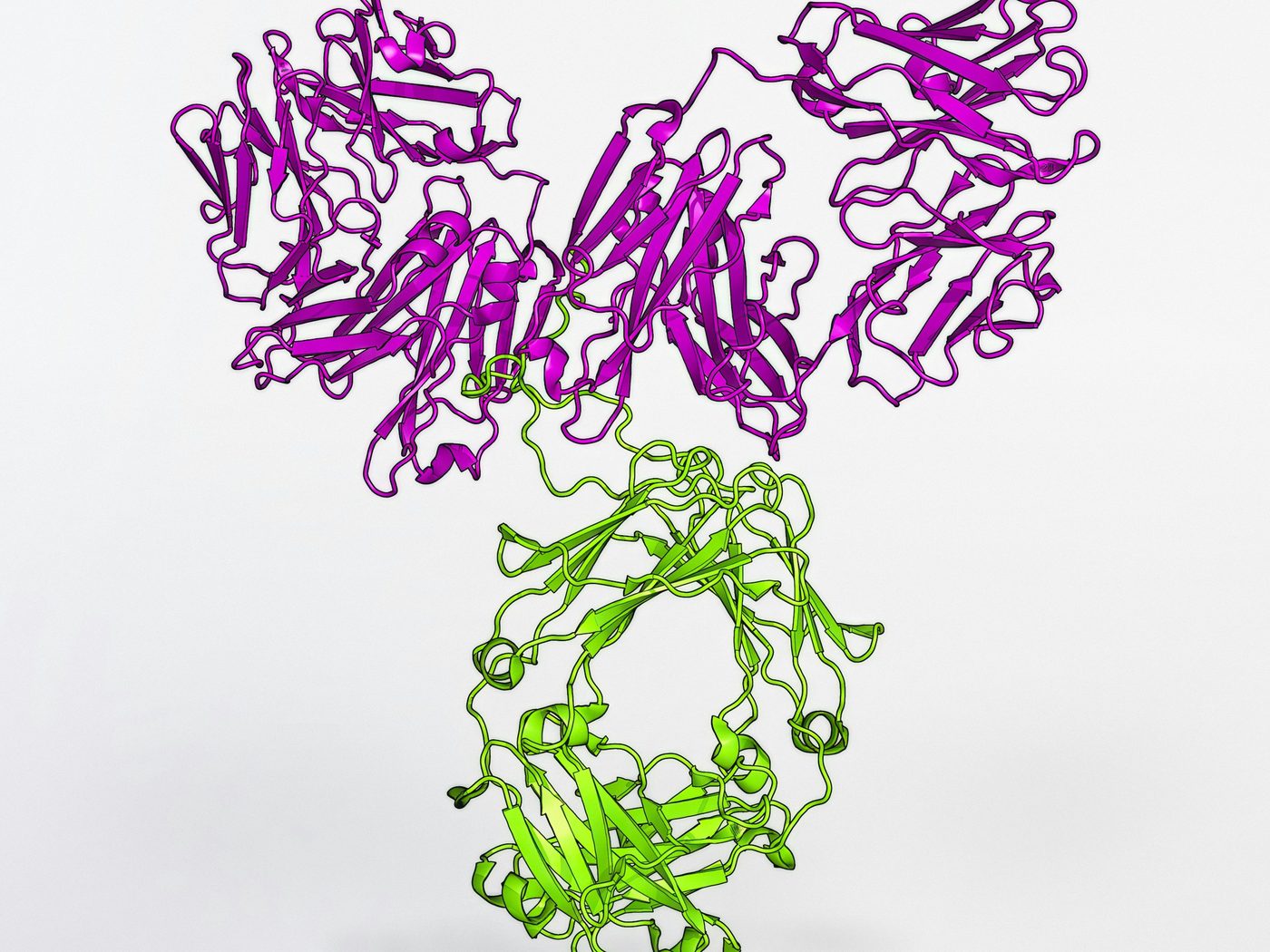
Build a molecule that grabs it. Lecanemab is a lab-made antibody, a Y-shaped immune protein, that binds tightly to the soluble protofibrils.
Measure two things in patients. Does the amyloid go down on PET brain scans, and do memory and daily function decline more slowly?
It's one strategy among several: NIA also describes roles for tau, the brain's immune cells and blood vessels.
A ribbon model of an antibody called VRC01, not lecanemab. Lecanemab is an antibody of the same Y-shaped kind, designed to grab amyloid-beta.
Lecanemab: what one large trial found
The key study is the Clarity AD trial, published by van Dyck and colleagues in the New England Journal of Medicine (online November 2022, in print January 2023). It enrolled 1,795 people aged 50 to 90 with early Alzheimer's, meaning mild cognitive impairment or mild dementia, and a PET scan or spinal fluid test showing amyloid. Half were randomly assigned to lecanemab by IV drip every two weeks, half to a placebo.
Both groups declined. On a clinical rating called CDR-SB, which runs from 0 to 18 (higher means more impairment), scores rose over 18 months by 1.21 points with lecanemab and 1.66 with placebo. The gap of 0.45 points is about 27% of the placebo group's decline. That's where the "27% slower" figure you may have seen comes from. PET scans in a substudy of 698 people also showed less brain amyloid with lecanemab.
The US Food and Drug Administration (FDA) gave lecanemab (brand name Leqembi) accelerated approval in January 2023 and traditional approval on July 6, 2023, for the same early stage.
Now the limits. Lecanemab slows decline; it doesn't stop or reverse it. The 27% is a relative number on one scale, and the actual gap was less than half a point out of 18. The FDA called the slowing clinically meaningful, while an editorial published with the trial called the effects "modest."
There are real risks, too. Reactions to the IV drip affected 26.4% of people on lecanemab, and 12.6% had brain swelling seen on scans, which usually goes away. This swelling, sometimes with small spots of bleeding, is called ARIA (amyloid-related imaging abnormalities). It often causes no symptoms but can be serious, even fatal, so the label carries a boxed warning. The current label also asks for brain MRI scans before treatment and after 1, 2, 3 and 6 months. The risk is higher in people with two copies of the ApoE ε4 gene variant.
Lecanemab is given through an IV drip. In the Clarity AD trial, 26.4% of people on the drug had a reaction to the infusion.
Try it on a laptop: four steps with a free fibril structure
You can't run a drug trial, but you can study the 3D shape of one form of amyloid-beta clump, shared free in the Protein Data Bank (PDB). Plan on six to eight weeks at a few hours a week, including two weeks of writing. No people are involved, so you won't need ethics review or consent forms.
Step 1. Pick one protein: amyloid-beta 42 (about 1 week)
Read NIA's page on what happens to the brain in Alzheimer's and the short PDB-101 article "Molecule of the Month: Amyloids." Then get the sequence. In UniProt entry P05067, amyloid-beta 42 is positions 672 to 713 of the amyloid precursor protein: 42 letters starting DAEFRHDSGY.
Step 2. Download PDB 5OQV, free (one afternoon)
PDB entry 5OQV is an amyloid-beta 42 fibril solved by cryo-electron microscopy (imaging frozen samples with electrons), published by Gremer and colleagues in Science in 2017. Its resolution is 4.0 Å (one ångström is a ten-billionth of a meter). Open it in the free Mol* viewer at rcsb.org/3d-view/5OQV, in your browser, or save the file from the entry page. PDBj, the worldwide PDB's partner site in Japan, has the same entry and a Korean-language menu.
Step 3. Find where the strands stack (1 to 2 weeks)
Color the model by chain. You'll see nine copies of the 42-amino-acid chain, each folded into what the authors call an "LS" shape, in two side-by-side stacks called protofilaments: chains A, C, E, F and H in one, B, D, G and I in the other. (The full fibril twists, but too slowly to see here.)
In Mol*, turn on Selection Mode and set the Picking Level to atom. Click the same atom, such as the backbone atom labeled CA in Phe19, in chains A and C, which sit directly on top of each other. Then choose Add, then Distance, in the Measurements panel. You should get about 4.7 Å, matching the 4.67 Å rise between layers reported in the paper. Next, list the amino acids pointing into the core and check them against the three water-avoiding (hydrophobic) clusters the authors name.
Step 4. Ask why the clumps form (2 to 3 weeks)
Paste the sequence into Expasy ProtScale and choose the Kyte & Doolittle scale, which scores how strongly each amino acid avoids water. The default window of 9 skips the first and last four positions of this short peptide, so also try window size 3. Do the peaks match the buried residues you found? Write down any that don't. Then mark positions 21 to 23, where Gremer's team notes five disease-causing inherited mutations, in a region that forms part of the fibril's outer edges.
Name the limits in your write-up. 5OQV is a snapshot of a finished fibril grown in a test tube under acidic conditions, so it can't show how clumping begins. In 2022, Yang and colleagues reported that amyloid-beta 42 filaments from human brains, such as PDB 7Q4B, differ from lab-made ones; 7Q4B's ordered core starts at residue 9. And since lecanemab binds protofibrils, a different form, your project describes the clump's shape, not how the drug works.
Your write-up can be a poster or a 4- to 6-page paper with three figures: the fibril with your measured spacing, the hydropathy plot with buried residues marked, and 5OQV next to 7Q4B. List every tool and setting in your methods section so someone else can repeat your work.
Three questions for one fibril file
For a laptop project like this one, a good first question has a clear method, public data and an answer you can show in one figure.
Too big
Workable on a laptop
How do we stop amyloid from clumping?
Are the residues buried in 5OQV's core the most water-avoiding ones on the Kyte & Doolittle scale?
Is the lab-grown fibril the same as the one in patients?
Which core residues do 5OQV and the brain-derived fibril 7Q4B share, and which differ?
Why do inherited mutations in amyloid-beta cause disease?
Do positions 21 to 23 sit on the outer edge in 7Q4B, as they do in 5OQV?
None of these will explain Alzheimer's, and that's fine. Each is a first step you can finish and check.
Reviewing the debate, or proposing the next study
The fibril file is one way into this topic. A literature review is another: you read published studies and compare what they found. You could collect the papers that debate the lecanemab result and ask how researchers decide whether a 0.45-point gap on an 18-point scale is big enough for patients and families to notice. PubMed and Google Scholar hold the published papers, and ClinicalTrials.gov lets you look up each trial by name.
A research proposal is the place for a study that needs patients, scans and lab tests. Your question might be whether a blood test could replace the PET scan or spinal fluid test that Clarity AD used to confirm amyloid. Your method: give both tests to the same patients at memory clinics and compare the results. You'd also list the samples and scans you'd need, and the ethics board approval and patient consent any study with people requires.
Starting with one protein
Her story was the question. Now research it.
CRI students work on questions of their own with professors in many fields, such as biology, economics, chemistry and history, and each student writes their own paper, whether that's original research, a literature review or a research proposal. If a project like this appeals to you, take a look at CRI's research programs.
Sources
National Institute on Aging (2024). What happens to the brain in Alzheimer's disease? National Institutes of Health. link
National Institute on Aging (2023). Alzheimer's disease fact sheet. National Institutes of Health. link
National Institute on Aging (2023). Memory problems, forgetfulness, and aging. National Institutes of Health. link
van Dyck, C. H., et al. (2023). Lecanemab in early Alzheimer's disease. New England Journal of Medicine, 388(1), 9-21 (published online November 29, 2022). link
Gandy, S., & Ehrlich, M. E. (2023). Moving the needle on Alzheimer's disease with an anti-oligomer antibody. New England Journal of Medicine, 388(1), 80-81. link
U.S. Food and Drug Administration (2023, July 6). FDA converts novel Alzheimer's disease treatment to traditional approval. link
Eisai Inc. (2026). Leqembi (lecanemab) prescribing information. DailyMed, U.S. National Library of Medicine. link
Gremer, L., et al. (2017). Fibril structure of amyloid-β(1-42) by cryo-electron microscopy. Science, 358(6359), 116-119. link
RCSB Protein Data Bank. Entry 5OQV: Near-atomic resolution fibril structure of complete amyloid-beta(1-42) by cryo-EM. link
Yang, Y., et al. (2022). Cryo-EM structures of amyloid-β 42 filaments from human brains. Science, 375(6577), 167-172. link
RCSB Protein Data Bank. Entry 7Q4B: Type I beta-amyloid 42 filaments from human brain. link
Goodsell, D. (2015). Molecule of the Month: Amyloids. PDB-101, RCSB Protein Data Bank. link
UniProt. Entry P05067: Amyloid-beta precursor protein. link
Mol* Viewer documentation. Making selections (Selection Mode and Picking Level). link
Mol* Viewer documentation. Managing the display (Measurements panel). link
SIB Swiss Institute of Bioinformatics. ProtScale. Expasy. link